| Betz cell | |
|---|---|
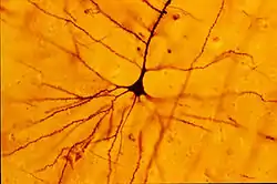
 A human neocortical pyramidal neuron such as a Betz Cell stained via Golgi technique. | |
| Details | |
| Location | Layer V of Cortex in primary motor cortex |
| Shape | Multipolar Pyramidal -- some of the longest axons in the body. |
| Function | excitatory projection neuron to spinal cord |
| Neurotransmitter | Glutamate |
| Presynaptic connections | Superficial cortical layers, premotor cortex |
| Postsynaptic connections | Ventral horn of the spinal cord |
| Identifiers | |
| NeuroLex ID | sao786552500 |
| Anatomical terms of neuroanatomy | |
Betz cells (also known as pyramidal cells of Betz) are giant pyramidal cells (neurons) located within the fifth layer of the grey matter in the primary motor cortex. These neurons are the largest in the central nervous system, sometimes reaching 100 μm in diameter.[1][2]
Betz cells are upper motor neurons that send their axons down to the spinal cord via the corticospinal tract, where in humans they synapse directly with anterior horn cells, which in turn synapse directly with their target muscles. Betz cells are not the sole source of direct connections to those neurons because most of the direct corticomotorneuronal cells are medium or small neurons.[3] While Betz cells have one apical dendrite typical of pyramidal neurons, they have more primary dendritic shafts, which can branch out at almost any point from the soma (cell body).[4] These perisomatic (around the cell body) and basal dendrites project into all cortical layers, but most of their horizontal branches/arbors populate layers V and VI, some reaching down into the white matter.[5] According to one study, Betz cells represent about 10% of the total pyramidal cell population in layer Vb of the human primary motor cortex.[6]
Betz cells are named after Ukrainian scientist Volodymyr Betz, who described them in his work published in 1874.[7]
See also
Notes
- ↑ Purves, Dale; George J. Augustine; David Fitzpatrick; William C. Hall; Anthony-Samuel LaMantia; James O. McNamara & Leonard E. White (2008). Neuroscience (4th ed.). Sinauer Associates. pp. 432–4. ISBN 978-0-87893-697-7.
- ↑ Nolte, J. The Human Brain, 5th ed. Mosby: Missouri; 2002, p.527. ISBN 0-323-01320-1
- ↑ Strick, Peter L.; Dum, Richard P.; Rathelot, Jean-Alban (8 July 2021). "The Cortical Motor Areas and the Emergence of Motor Skills: A Neuroanatomical Perspective". Annual Review of Neuroscience. 44 (1): 425–447. doi:10.1146/annurev-neuro-070918-050216. PMID 33863253. S2CID 233278281.
- ↑ Braak, H; Braak, E (1976). "The pyramidal cells of Betz within the cingulate and precentral gigantopyramidal field in the human brain. A Golgi and pigmentarchitectonic study". Cell and Tissue Research. 172 (1): 103–19. doi:10.1007/bf00226052. PMID 991201. S2CID 40242681.
- ↑ Meyer, G (1987). "Forms and spatial arrangement of neurons in the primary motor cortex of man". The Journal of Comparative Neurology. 262 (3): 402–28. doi:10.1002/cne.902620306. PMID 3655019. S2CID 45950277.
- ↑ Rivara, CB; Sherwood, CC; Bouras, C; Hof, PR (2003). "Stereologic characterization and spatial distribution patterns of Betz cells in the human primary motor cortex". The Anatomical Record Part A: Discoveries in Molecular, Cellular, and Evolutionary Biology. 270 (2): 137–51. doi:10.1002/ar.a.10015. PMID 12524689.
- ↑ Betz W. (1874) Anatomischer Nachweis zweier Gehirncentra. Centralblatt für die medizinischen Wissenschaften. 12:578-580, 595-599.