| Abbreviation | ER-C |
|---|---|
| Established | 27 January 2004 |
| Type | Institute within Forschungszentrum Jülich and User Facility jointly operated with RWTH Aachen |
| Location | |
| Coordinates | 50°54′29″N 6°24′49″E / 50.90806°N 6.41361°E |
Directors | Rafal E. Dunin-Borkowski, Joachim Mayer and Carsten Sachse |
Founding Director | Knut Urban |
| Affiliations | Jülich Research Centre and RWTH Aachen University |
Staff | 50-100 |
| Website | www |
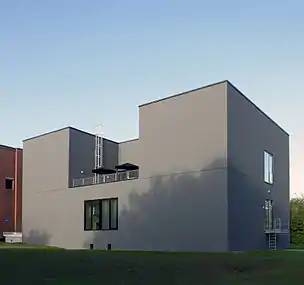
The Ernst Ruska-Centre for Microscopy and Spectroscopy with Electrons (ER-C) is an institute located on the campus of Forschungszentrum Jülich belonging to the Helmholtz Association of German Research Centres. It comprises three divisions: ER-C-1 “Physics of Nanoscale systems”, ER-C-2 “Materials Science and Technology” and ER-C-3 “Structural Biology”.
Within the framework of a competence platform governed jointly by Forschungszentrum Jülich and RWTH Aachen University, the ER-C operates a national and international user facility that provides access to state-of-the-art instruments, methods and expertise to universities, research institutions and industry.
The ER-C's main purposes are fundamental research in electron microscopy, focusing on method development and applications of high-resolution transmission electron microscopy (HRTEM) and scanning-transmission electron microscopy (STEM) in physics, chemistry and biology.
History
As a competence platform, the ER-C was founded on 27 January 2004 through a contract signed by the chairman of Forschungszentrum Jülich Joachim Treusch and the rector of RWTH Aachen University Burkhard Rauhut.[1] It was inaugurated on 18 May 2006 in the presence of members of the Ernst Ruska family, as well as representatives of the international electron microscopy community.[2] On 1 January 2017, the ER-C attained the status of an independent scientific institute in Forschungszentrum Jülich. The ER-C is presently expanding further within the framework of the Research Infrastructure Roadmap of the German Federal Ministry of Education and Research (BMBF) under the designation ER-C 2.0. The ER-C thus creates incentives for companies dealing with novel materials and technologies to settle in the Rhenish mining area and contribute to the development of a competence region for innovative materials technologies and ultimately to the success of structural change.
Instrumental Resources

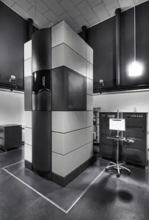
The ER-C develops new methods and technologies in the field of electron microscopy, with a special focus on ultra-high-resolution techniques to study solid state materials, soft materials and biological systems. The ER-C houses conventional and state-of-the-art electron microscopes, ranging from standard scanning electron microscopes to highly-specialised aberration corrected instruments offering sub-Å resolution imaging and spectroscopy,[3] as well as quantitative measurements of electromagnetic field distributions using phase contrast techniques that include off-axis electron holography and 4D STEM. The ER-C currently operates seven aberration-corrected instruments.[4]
On 29 February 2012, the ER-C inaugurated the first chromatic aberration corrected transmission electron microscope in Europe, which is designated “PICO” and is capable of resolving atomic positions in materials with a spatial resolution of 50 picometers and a precision approaching 1 picometer.[5] It is also equipped with a monochromator, an electron biprism, an electron energy-loss spectrometer and a direct electron counting detector.
In situ and quantitative electromagnetic field measurements can be carried out using a spherical aberration corrected transmission electron microscope equipped with a large (11 mm) objective pole-piece gap, a double biprism system and a direct electron counting detector. The same microscope is used for ongoing instrumentation development, including ultra-high vacuum sample transfer, laser illumination, in situ magnetising and low temperature experiments.
Recently, cryo-electron microscopy (cryo-EM) became an integral part of the Ernst-Ruska Centre with state-of-the-art cryo-microscopes: 300 kV Titan Krios G4 (operational in Summer 2021) and 200 kV Talos Arctica including Gatan Bioquantum K3 detectors.[6]
Research Programmes

The three scientific divisions of the ER-C focus on specific research topics:
1. ERC-1 (solid state physics and chemistry) focuses on electroceramics and nanoelectronic oxides, materials for Green IT, electromagnetic field mapping, electron optics and method development, metallic alloys and crystal growth, catalysis, nanofabrication and quantum sensing, scanning tunneling microscopy and spectroscopy and momentum-resolved scanning transmission electron microscopy.
2. ERC-2 (materials science) focuses on gas separation membranes, battery materials, non-volatile memories and high-performance steels.
3. ERC-3 (structural biology) focuses on cryoelectron microscopy (cryo-EM) using a comprehensive single-particle and tomography approach to investigate the structures of membrane biology processes autophagy and endocytosis.
The facilities are accessible to external users through the ER-C user office.
References
- ↑ "Forschungszentrum Jülich - Press releases - Imaging the World of Atoms". www.fz-juelich.de. Retrieved 2021-03-22.
- ↑ "Forschungszentrum Jülich - Press releases - Looking at atoms through the eyes of TITANS". www.fz-juelich.de. Retrieved 2021-03-22.
- ↑ "Forschungszentrum Jülich - Press releases - Work Begins on Laboratory for World's Most Powerful Microscope". www.fz-juelich.de. Retrieved 2021-03-22.
- ↑ "Forschungszentrum Jülich - Ernst Ruska-Centre for Microscopy and Spectroscopy with Electrons (ER‑C)". www.fz-juelich.de. Retrieved 2021-03-22.
- ↑ Press Release: Electron Microscopy Enters the Picometre Scale
- ↑ "Forschungszentrum Jülich - Structural Biology (ER-C-3)". www.fz-juelich.de. Retrieved 2021-03-22.