| H1-2 | |||||||||||||||||||||||||||||||||||||||||||||||||||
|---|---|---|---|---|---|---|---|---|---|---|---|---|---|---|---|---|---|---|---|---|---|---|---|---|---|---|---|---|---|---|---|---|---|---|---|---|---|---|---|---|---|---|---|---|---|---|---|---|---|---|---|
| Identifiers | |||||||||||||||||||||||||||||||||||||||||||||||||||
| Aliases | H1-2, H1.2, H1C, H1F2, H1s-1, histone cluster 1, H1c, histone cluster 1 H1 family member c, HIST1H1C, H1.2 linker histone, cluster member | ||||||||||||||||||||||||||||||||||||||||||||||||||
| External IDs | OMIM: 142710 MGI: 1931526 HomoloGene: 68455 GeneCards: H1-2 | ||||||||||||||||||||||||||||||||||||||||||||||||||
| |||||||||||||||||||||||||||||||||||||||||||||||||||
| |||||||||||||||||||||||||||||||||||||||||||||||||||
| |||||||||||||||||||||||||||||||||||||||||||||||||||
| |||||||||||||||||||||||||||||||||||||||||||||||||||
| |||||||||||||||||||||||||||||||||||||||||||||||||||
| Wikidata | |||||||||||||||||||||||||||||||||||||||||||||||||||
| |||||||||||||||||||||||||||||||||||||||||||||||||||
Histone H1.2 is a protein that in humans is encoded by the HIST1H1C gene.[5][6][7]
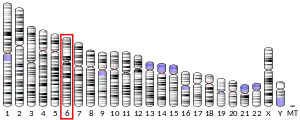
Histones are basic nuclear proteins responsible for nucleosome structure of the chromosomal fiber in eukaryotes. Two molecules of each of the four core histones (H2A, H2B, H3, and H4) form an octamer, around which approximately 146 bp of DNA is wrapped in repeating units, called nucleosomes. The linker histone, H1, interacts with linker DNA between nucleosomes and functions in the compaction of chromatin into higher order structures. This gene is intronless and encodes a member of the histone H1 family. Transcripts from this gene lack polyA tails but instead contain a palindromic termination element. This gene is found in the large histone gene cluster on chromosome 6.[7]
Apart from its roles in the nucleus, histone H1.2 also participates in apoptosis. In response to apoptotic stimuli, mainly DNA damage, it is translocated from the nucleus to the cytosol. There, it activates Bak, a pro-apoptotic protein bound to the mithochondria outer membrane (MOM). Activation of Bak causes the perforation of the mitochondria, a process known as MOMP (mitochondria outer membrane permeabilization) which promotes apoptosis. Histone H1.2 also forms a complex with the apoptosome, possibly regulating its formation.
References
- 1 2 3 GRCh38: Ensembl release 89: ENSG00000187837 - Ensembl, May 2017
- 1 2 3 GRCm38: Ensembl release 89: ENSMUSG00000036181 - Ensembl, May 2017
- ↑ "Human PubMed Reference:". National Center for Biotechnology Information, U.S. National Library of Medicine.
- ↑ "Mouse PubMed Reference:". National Center for Biotechnology Information, U.S. National Library of Medicine.
- ↑ Eick S, Nicolai M, Mumberg D, Doenecke D (Sep 1989). "Human H1 histones: conserved and varied sequence elements in two H1 subtype genes". Eur J Cell Biol. 49 (1): 110–5. PMID 2759094.
- ↑ Marzluff WF, Gongidi P, Woods KR, Jin J, Maltais LJ (Oct 2002). "The human and mouse replication-dependent histone genes". Genomics. 80 (5): 487–98. doi:10.1016/S0888-7543(02)96850-3. PMID 12408966.
- 1 2 "Entrez Gene: HIST1H1C histone cluster 1, H1c".
Further reading
- Ohe Y, Hayashi H, Iwai K (1990). "Human spleen histone H1. Isolation and amino acid sequences of three minor variants, H1a, H1c, and H1d". J. Biochem. 106 (5): 844–57. doi:10.1093/oxfordjournals.jbchem.a122941. PMID 2613692.
- Eilers A, Bouterfa H, Triebe S, Doenecke D (1994). "Role of a distal promoter element in the S-phase control of the human H1.2 histone gene transcription". Eur. J. Biochem. 223 (2): 567–74. doi:10.1111/j.1432-1033.1994.tb19026.x. PMID 8055927.
- Albig W, Drabent B, Kunz J, et al. (1993). "All known human H1 histone genes except the H1(0) gene are clustered on chromosome 6". Genomics. 16 (3): 649–54. doi:10.1006/geno.1993.1243. PMID 8325638.
- Albig W, Doenecke D (1998). "The human histone gene cluster at the D6S105 locus". Hum. Genet. 101 (3): 284–94. doi:10.1007/s004390050630. PMID 9439656. S2CID 38539096.
- Richardson RT, Batova IN, Widgren EE, et al. (2000). "Characterization of the histone H1-binding protein, NASP, as a cell cycle-regulated somatic protein". J. Biol. Chem. 275 (39): 30378–86. doi:10.1074/jbc.M003781200. PMID 10893414.
- Parseghian MH, Newcomb RL, Winokur ST, Hamkalo BA (2001). "The distribution of somatic H1 subtypes is non-random on active vs. inactive chromatin: distribution in human fetal fibroblasts". Chromosome Res. 8 (5): 405–24. doi:10.1023/A:1009262819961. PMID 10997781. S2CID 28425496.
- Strausberg RL, Feingold EA, Grouse LH, et al. (2003). "Generation and initial analysis of more than 15,000 full-length human and mouse cDNA sequences". Proc. Natl. Acad. Sci. U.S.A. 99 (26): 16899–903. Bibcode:2002PNAS...9916899M. doi:10.1073/pnas.242603899. PMC 139241. PMID 12477932.
- Gerhard DS, Wagner L, Feingold EA, et al. (2004). "The status, quality, and expansion of the NIH full-length cDNA project: the Mammalian Gene Collection (MGC)". Genome Res. 14 (10B): 2121–7. doi:10.1101/gr.2596504. PMC 528928. PMID 15489334.
- Garcia BA, Busby SA, Barber CM, et al. (2005). "Characterization of phosphorylation sites on histone H1 isoforms by tandem mass spectrometry". J. Proteome Res. 3 (6): 1219–27. doi:10.1021/pr0498887. PMID 15595731.
- Andersen JS, Lam YW, Leung AK, et al. (2005). "Nucleolar proteome dynamics". Nature. 433 (7021): 77–83. Bibcode:2005Natur.433...77A. doi:10.1038/nature03207. PMID 15635413. S2CID 4344740.
- Duce JA, Smith DP, Blake RE, et al. (2006). "Linker histone H1 binds to disease associated amyloid-like fibrils". J. Mol. Biol. 361 (3): 493–505. doi:10.1016/j.jmb.2006.06.038. PMID 16854430.
- Olsen JV, Blagoev B, Gnad F, et al. (2006). "Global, in vivo, and site-specific phosphorylation dynamics in signaling networks". Cell. 127 (3): 635–48. doi:10.1016/j.cell.2006.09.026. PMID 17081983. S2CID 7827573.


