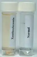
| Xanthochromia | |
|---|---|
 |
Xanthochromia, from the Greek xanthos (ξανθός) "yellow" and chroma (χρώμα) "colour", is the yellowish appearance of cerebrospinal fluid that occurs several hours after bleeding into the subarachnoid space caused by certain medical conditions, most commonly subarachnoid hemorrhage.[1] Its presence can be determined by either spectrophotometry (measuring the absorption of particular wavelengths of light) or simple visual examination. It is unclear which method is superior.[2]
Physiology
Cerebrospinal fluid, which fills the subarachnoid space between the arachnoid membrane and the pia mater surrounding the brain, is normally clear and colorless. When there has been bleeding into the subarachnoid space, the initial appearance of the cerebrospinal fluid can range from barely tinged with blood to frankly bloody, depending on the extent of bleeding. Within several hours, the red blood cells in the cerebrospinal fluid are destroyed, releasing their oxygen-carrying molecule heme, which is then metabolized by enzymes to bilirubin, a yellow pigment. The most common cause for bleeding into the subarachnoid space is a subarachnoid hemorrhage from a ruptured cerebral aneurysm.[3]
The most frequently employed initial test for subarachnoid hemorrhage is a computed tomography scan of the head, but it detects only 98% of cases in the first 12 hours after the onset of symptoms, and becomes less useful afterwards.[4] Therefore, a lumbar puncture ("spinal tap") is recommended to obtain cerebrospinal fluid if someone has symptoms of a subarachnoid hemorrhage (e.g., a thunderclap headache, vomiting, dizziness, new-onset seizures, confusion, a decreased level of consciousness or coma, neck stiffness or other signs of meningismus, and signs of sudden elevated intracranial pressure), but no blood is visible on the CT scan.[1] According to one article, a spinal tap is not necessary if no blood is seen on a CT scan done using a third generation scanner within six hours of the onset of the symptoms. However, this is not standard of care.[5][6]
Heme from red blood cells (RBC) that are in the cerebrospinal fluid because a blood vessel was damaged during the lumbar puncture (a "traumatic tap") has no time to be metabolized, and therefore no bilirubin is present.
After the cerebrospinal fluid is obtained, a variety of its parameters can be checked, including the presence of xanthochromia. If the cerebrospinal fluid is bloody, it is centrifuged to determine its color.
Spectrophotometry
Many laboratories rely on only the color of the cerebrospinal fluid to determine the presence or absence of xanthochromia.[7] However, recent guidelines suggest that spectrophotometry should be performed. Spectrophotometry relies on the different transmittance, or conversely, absorbance, of light by different substances or materials, including solutes.[8] Bilirubin absorbs light at wavelengths between 450 and 460 nm.[9] Spectrophotometry can also detect the presence of oxyhemoglobin and methemoglobin, which absorb light at 410-418 nm and 403-410 nm, respectively, and also may indicate that bleeding has occurred; to identify substances in cerebrospinal fluid that absorb light at other wavelengths but are not due to bleeding, such as carotenoids;[1][7] and to detect very small amounts of yellow color saturation (about 0.62%) which may be missed by visual inspection, especially when the cerebrospinal fluid has been examined under incandescent lighting or a tungsten desk lamp (corresponding to International Commission on Illumination standard illuminant A).[8]
Visual inspection is the most frequent method used in the United States to assess cerebrospinal fluid for xanthochromia,[10] while spectrophotometry is used on up to 94% of specimens in the United Kingdom.[1][9] There is still disagreement about whether or not to routinely use spectrophotometry or whether visual inspection is adequate, and one group of authors has even advocated measuring bilirubin levels.[11]
See also
References
- 1 2 3 4 Cruickshank, A; Auld, P.; Beetham, R.; et al. (May 2008). "Revised National Guidelines for Analysis of Cerebrospinal Fluid for Bilirubin in Suspected Subarachnoid Haemorrhage". Annals of Clinical Biochemistry. 45 (Pt 3): 238–244. doi:10.1258/acb.2008.007257. PMID 18482910. S2CID 24393459.
- ↑ Chu, K; Hann, A; Greenslade, J; Williams, J; Brown, A (Mar 10, 2014). "Spectrophotometry or Visual Inspection to Most Reliably Detect Xanthochromia in Subarachnoid Hemorrhage: Systematic Review". Annals of Emergency Medicine. 64 (3): 256–264.e5. doi:10.1016/j.annemergmed.2014.01.023. PMID 24635988.
- ↑ van Gijn, J.; Kerr, R.S.; Rinkel, G.J. (2007). "Subarachnoid Haemorrhage". Lancet. 369 (9558): 306–318. doi:10.1016/S0140-6736(07)60153-6. PMID 17258671. S2CID 29126514.
- ↑ van der Wee, N.; Rinkel, G.J.; Hasan, D.; van Gijn J., J. (March 1995). "Detection of Subarachnoid Haemorrhage on Early CT: Is Lumbar Puncture Still Needed After a Negative Scan?". Journal of Neurology, Neurosurgery, and Psychiatry. 58 (3): 357–359. doi:10.1136/jnnp.58.3.357. PMC 1073376. PMID 7897421.
- ↑ Perry, J.J.; Stiell, I.G.; Sivilotti, M.L.; et al. (July 2011). "Sensitivity of Computed Tomography Performed Within Six Hours of Onset of Headache for Diagnosis of Subarachnoid Haemorrhage: Prospective Cohort Study". British Medical Journal. 343: d4277. doi:10.1136/bmj.d4277. PMC 3138338. PMID 21768192.
- ↑ Stewart, H.; Reuben, A.; McDonald, J. (September 2014). "LP or not LP, That is the Question: Gold Standard or Unnecessary Procedure in Subarachnoid Haemorrhage?". Emergency Medicine Journal. 31 (9): 720–723. doi:10.1136/emermed-2013-202573. PMID 23756363. S2CID 206934433.
- 1 2 Edlow, J.A. (July 2004). "The Worst Headache". Morbidity & Mortality Rounds on the Web. Agency for Healthcare Research and Quality. Retrieved 2008-06-22.
- 1 2 Williams, Anna (2004). "Xanthochromia in the cerebrospinal fluid" (PDF). Practical Neurology. 126 (4): 174–175.
- 1 2 Petzold, Axel; Keir, Geoffrey; Sharpe, Lindsay T. (2004). "Spectrophotometry for Xanthochromia". New England Journal of Medicine. 351 (16): 1695–1696. doi:10.1056/nejm200410143511627. PMID 15483297.
- ↑ Edlow, J.A.; Bruner, K.S.; Horowitz, G.L. (April 2002). "Xanthochromia". Archives of Pathology and Laboratory Medicine. 126 (4): 413–415. doi:10.5858/2002-126-0413-X. PMID 11900563.
- ↑ Florkowski, Christopher; Ungerer, Jacobus; Southby, Sandi; George, Peter (17 December 2004). "CSF Bilirubin Measurement for Xanthochromia" (PDF). Journal of the New Zealand Medical Association. 117 (1207): U1231. PMID 15608818.