| Colpodella | |
|---|---|
 | |
| Scientific classification | |
| Domain: | Eukaryota |
| Clade: | Diaphoretickes |
| Clade: | TSAR |
| Clade: | SAR |
| Clade: | Alveolata |
| Phylum: | Apicomplexa |
| Order: | Colpodellida |
| Family: | Colpodellidae Simpson & Patterson, 1996 |
| Genus: | Colpodella Cienkowski, 1865 |
| Species | |
|
See text. | |
| Synonyms | |
| |
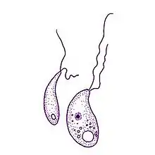
Colpodella is a genus of alveolates comprising 5 species, and two further possible species:[1] They share all the synapomorphies of apicomplexans, but are free-living, rather than parasitic.[1] Many members of this genus were previously assigned to a different genus - Spiromonas.
The type species is Colpodella pugnax Cienkowski 1865.
Description
These are small (< 20 μm in diameter) flagellated protists. The life cycle of consists of two main stages: flagellated trophozoites and cysts, which are the reproductive stage in the life cycle.
Morphologically the trophozoites of Colpodella are similar to Perkinsus zoospores, although the two taxa are not specifically related. The motile stages of both genera have a pair of anterior orthogonal flagella, vesicular mitochondrial cristae, inner alveolar membranes and micropores. Both Colpodella and Perkinsus species have open sided truncated conoids (sometimes called pseudoconoids), rhoptries that occupy the length of the cell and smaller micronemes. Both the rhoptries and micronemes arise at the anterior portion of the cell. A three-layered pellicle lies beneath the plasma membrane and is otherwise composed of the alveolar membranes and widely separated microtubules that arise subapically. Some species have extrusive organelles (trichocysts).
Unlike Perkinsus, Colpodella are free-living and are voracious predators of other free-living protists. Most species apparently penetrate through the cell membrane and consume the prey's cytoplasm - this mode of feeding is known as myzocytosis. While feeding the predator attaches its anterior portion - the rostrum - to the prey. The rostrum contains the pseudoconoid, which transforms into a ring of microtubules encircling the attachment zone. The cytoplasm of the prey is then drawn into a large posterior food vacuole.
Following feeding cells lose their flagella, become spherical, encyst and divide (i.e. reproduce). The cysts are simple spheres. The food vacuole appears as a large central vacuole in the cyst; as division progresses the remnant vacuole material is reduced to a residual body. Typically Colpodella divides into four daughter cells (sometimes just two).[1] This is in contrast to true Apicomplexa and Perkinsus, which typically produce many more daughter cells during reproduction - Perkinsus species can produce up to 32 cells, for example, while Toxoplasma produces 128. The daughter cells grow flagella, the cyst wall ruptures, and the cells swim away, leaving the residual body behind. A possible sexual process has been observed in at least two species.[1]
Taxonomy
This family appears to be a sister clade to the Apicomplexa.[2] Their life style may be representative of the free living ancestors of the Apicomplexa. One significant difference is that this genus, like the Perkinsea, have an open sided conoid (pseudoconoid) while the Apicomplexa which possess a conoid (the Conoidasida) have a closed conoid.
Another genus in this family is Acrocoelus.
Species currently within genus:
- Colpodella edax (Klebs 1892) Simpson & Patterson 1996
- Colpodella pseudoedax Mylnikov & Mylnikov 2007
- Colpodella pugnax Cienkowsky 1865 non Simpson & Patterson 1996
- Colpodella angusta (Dujardin 1841) Simpson & Patterson 1996 [Dingensia angusta (Dujardin 1841) Patterson & Zoelffel 1991]
Species transferred to other genera:[3]
- Colpodella gonderi (Foissner & Foissner 1984) Simpson & Patterson 1996 as Microvorax gonderi (Foissner & Foissner 1984) Cavalier-Smith 2017
- Colpodella perforans (Hollande 1938) Patterson & Zölffel 1991 as Chilovora perforans (Hollande 1938) Cavalier-Smith & Chao 2004
- Colpodella pontica Mylnikov 2000 as Voromonas pontica (Mylnikov 2000) Cavalier-Smith & Chao 2004
- Colpodella pugnax Simpson & Patterson 1996 non Cienkowsky 1865 as Algovora pugnax (Simpson & Patterson 1996) Cavalier-Smith & Chao 2004
- Colpodella tetrahymenae Cavalier-Smith 2004 as Microvorax tetrahymenae (Cavalier-Smith & Chao 2004) Cavalier-Smith 2017
- Colpodella turpis Simpson & Patterson 1996 as Algovora turpis (Simpson & Patterson 1996) Cavalier-Smith & Chao 2004
- Colpodella unguis Patterson & Simpson 1996 as Colpovora unguis (Patterson & Simpson 1996) Cavalier-Smith 2017
- Colpodella vorax (Kent, 1880) Simpson & Patterson, 1996 as Dinomonas vorax Kent 1880
Clinical
These organisms are not normally considered to be human pathogens. However, a report of an infection of the erythrocytes in a Chinese woman with a deficiency of natural killer cells has been reported.[4]
References
- 1 2 3 4 Alastair G. B. Simpson; David J. Patterson (1996). "Ultrastructure and identification of the predatory flagellate Colpodella pugnax Cienkowski (Apicomplexa) with a description of Colpodella turpis n. sp. and a review of the genus". Systematic Parasitology. 33 (3): 187–198. doi:10.1007/BF01531200.
- ↑ Brian S. Leander; Olga N. Kuvardina; Vladimir V. Aleshin; Alexander P. Mylnikov; Patrick J. Keeling (2003). "Molecular phylogeny and surface morphology of Colpodella edax (Alveolata): insights into the phagotrophic ancestry of apicomplexans" (PDF). Journal of Eukaryotic Microbiology. 50 (5): 334–340. doi:10.1111/j.1550-7408.2003.tb00145.x. PMID 14563171. Archived from the original (PDF) on 2012-04-01. Retrieved 2011-10-14.
- ↑ Cavalier-Smith, Thomas (2018). "Kingdom Chromista and its eight phyla: a new synthesis emphasising periplastid protein targeting, cytoskeletal and periplastid evolution, and ancient divergences". Protoplasma. 255 (1): 297–357. doi:10.1007/s00709-017-1147-3. PMC 5756292. PMID 28875267.
- ↑ Yuan CL, Keeling PJ, Krause PJ, Horak A, Bent S, Rollend L, Hua XG (2012) Colpodella spp.-like parasite infection in woman, China. Emerg Infect Dis 18(1):125-127 doi: 10.3201/eid1801.110716
External links
- Leander, Brian S.; Keeling, Patrick J. (2003). "Morphostasis in alveolate evolution" (PDF). Trends in Ecology and Evolution. 18 (8): 395–402. CiteSeerX 10.1.1.452.8288. doi:10.1016/S0169-5347(03)00152-6. Archived from the original (PDF) on 2012-04-01. Retrieved 2011-10-14.